Ever wanted to hone your skills as a scientific sleuth? Now’s your chance.
Thanks to the American Society for Biochemistry and Molecular Biology (ASBMB), which is committed to educating authors on best practices in publishing, figure preparation, and reproducibility, we’re presenting the fifteenth in a series, Forensics Friday.
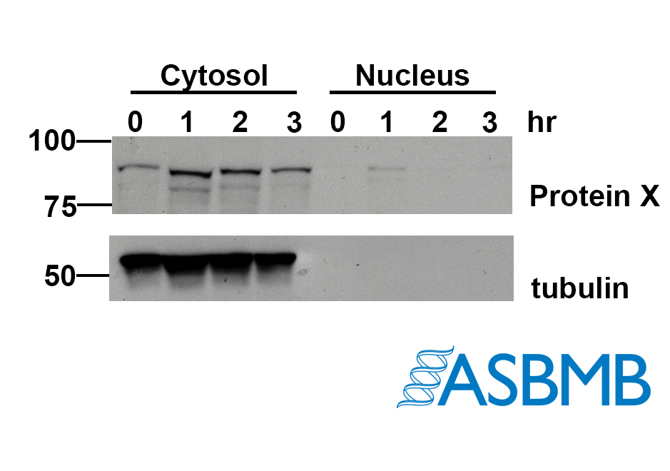
Take a look at the image below, and then take our poll. (We recommend using the Chrome browser.) After that, click on the link below to find out the right answer.

Think you chose the right answer? Click here to find out.
And check out the last Forensics Friday.
Like Retraction Watch? You can make a tax-deductible contribution to support our work, follow us on Twitter, like us on Facebook, add us to your RSS reader, sign up for an email every time there’s a new post (look for the “follow” button at the lower right part of your screen), or subscribe to our daily digest. If you find a retraction that’s not in our database, you can let us know here. For comments or feedback, email us at [email protected].
How can there be nothing wrong with this image, if the lanes for Protein X and tub don’t match neither in lane width nor running direction?
These were actually run on the same gel, but it’s hard to tell because the proteins differ in abundance and the tubulin itself is over-exposed.
but the lanes usually don’t get less broad towards the bottom of the gel. I reckon I would want to see the original scan.
Yes, agree with Chartinael. Tubulin band looks very odd.
Real question: aren’t they supposed to also have a nucleus specific protein / marker to indicate that their nucleus extract does not simply contain less material ?
Exactly! That’s what I would want to see if I were a reviewer!
That was my thought too. I’d like to see a nuclear loading control.
No way the bottom part is from the same gel, even assuming the strong signal.
The absence of tubulin in the cytosolic lanes suggests a good fractionation, but including a nuclear loading control (such as lamin or a histone-associated protein) would be much better. Would be up to the discretion of the referee whether this is a meaningful issue requiring additional data, and I imagine in plenty of cases this would slide through just fine as-is, but “there is nothing wrong” is rather debatable. Perhaps there’s “nothing wrong… with the image,” but IMO answer choice A is probably more correct.
This is where Forensics Friday falls over. Image adequacy and veracity is context dependent. Anyone reviewing a paper will have at least a few paragraphs of description of experiment aims and background to allow assessment of the adequacy of the image as a supporting piece of evidence. Please add such information in future.
A gel with a loading control band that shows nothing in some of the lanes on the face of things indicates there was little if any product inserted in those lanes. Having no information about how these cell fluids were obtained, during what phase of the cell cycle, leaves a reviewer in the dark as to whether observed issues with the image are reasonable.
Forensics Friday would be much more valuable with some description of what purportedly is being assessed, and the conditions involved. Some information about the type of assay producing the images is necessary.
Seeing nothing in a loading control band for some lanes leaves the reviewer with no idea as to whether seeing anything in other bands of those lanes means anything.
A competent reviewer would ask for additional material, not because there’s anything indicating image manipulation, but because absence of loading control information leaves interpretation of the other bands in those lanes up in the air. Statistical analysis of relative band densities requires loading control values for an adjusting covariate in models of such data.
A competent reviewer would not choose B.