 There’s something strange about a 2008 paper on the role of nicotine receptors in promoting lung cancer: One of the western blot analyses looks like a version of an image from a commercial catalog.
A commenter on PubPeer pointed out the similarities between an image in “Role of α7-nicotinic acetylcholine receptor in human non-small cell lung cancer proliferation,” which was published in Cell Proliferation, and one used to promote an enzyme sold by Cell Signaling Technology.
Unfortunately, if the images are indeed the same, we can’t tell for certain who copied from whom. But a representative of the company told us it generates its own images, and this one was likely created before the paper was published. The last author — for whom we’ve found three other retractions — denied that the paper copied the image.
Here’s the panel from the paper, which was published in 2008:
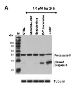
Characterization of apoptosis induced by d-tubocurarine, α-CbT, mutated-α-CbT or Erabutoxin-a on NSCLC cell line A549. Panel A: cleavage of procaspase-9. P
And here’s the catalog image — used to demonstrate the applications expression and activity of the caspase-9 enzyme — along with the description:
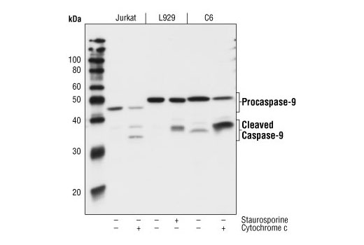
Western blot analysis of extracts from Jurkat cells (human), L929 cells (mouse), and C6 cells (rat), untreated or treated with staurosporine or cytochrome c as indicated, using Caspase 9 (C9) Mouse mAb.
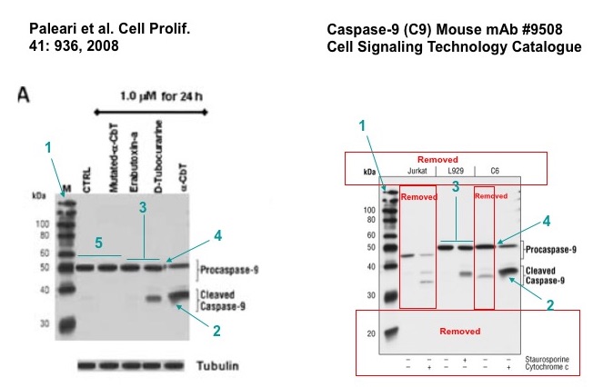
And here’s a side-by-side comparison, posted by someone on PubPeer:
A spokesperson for Cell Signaling Technology told us that their image “was originally published to our site in October of 2005” and they create their own images unless otherwise noted. He told us:
I can’t comment on any similarities or differences seen between the image in the paper and the image on our site but I can say that this is not the first time an image from our site has appeared in another location. Historically (2010-2013), may of our images were copied and displayed on other antibody vendors sites, we have submitted a number of C&D orders to a number of companies, mostly located abroad.
The spokesperson also said:
at this time we are keeping an eye on the situation but don’t feel it appropriate to take action.
The image issue isn’t the only problem with the paper, which has been cited 42 times, according to Thomson Reuters Web of Science. It has two corrections from 2009 — one pertaining to an incorrect panel in another figure, and one to an error in listing author affiliations. PubPeer commenters have suggested there may be other irregularities with images in the paper, as well.
Last author Patrizia Russo, who is currently affiliated with IRCCS San Raffaele Pisana, maintained that the images were different. She also told us that an Italian court has looked into potential wrong-doing in her work:
The Criminal Court of Genoa-Italy on 23.08.2013, through act n. 5243/13 RG GIP, Court Judge Dr. Bossi, filed and closed definitively the proceedings of any possible scientific misconduct.
She provided us with a copy of the court decision, which is in Italian.
This isn’t Russo’s first problem with a paper. She and the first author share two previous retractions. We reported on one a few years ago; The reason the retraction note gave was “concerns regarding the veracity of the data presented.” The other is from the International Journal of Cancer for a 2009 paper, “Inhibition of non-neuronal α7-nicotinic receptor reduces tumorigenicity in A549 NSCLC xenografts,” retracted “due to unverified data.”
We’ve found a third retraction for Russo: “Tumor necrosis factor enhances SN38-mediated apoptosis in mesothelioma cells: The role of nuclear factor-κB pathway activation,” published in 2005 and retracted in 2010 by Cancer, and cited 16 times. Here’s the retraction note:
On October 6, 2009, we were alerted to concerns about the integrity of the data in the article. A formal investigation was conducted by the Institutional Office for Research Integrity (UIR) at the National Institute for Cancer Research (IST) in Genoa, Italy. The investigation report from the UIR President, dated November 4, 2009, stated the following:
“1) The internal investigation is concluded and no further analyses are planned on the issue.
2) The UIR concluded that the article contains evidence of data fabrication/duplication.
3) All the authors (either belonging or not to the IST) have been informed of the concerns about data integrity. Some authors acknowledged the conclusion stated in the previous point 2, others did not; however, none of them accepted the responsibility of having fabricated the data.”
Based on this report and our concerns about the validity of the study, we hereby retract this article from Cancer and from the medical literature.
An email to the first author of the 2008 Cell Proliferation paper, Laura Paleari, listed as affiliated with the National Cancer Research Institute in Italy, has bounced back.
We contacted the editor in chief of Cell Proliferation, Catherine Sarraf, who told us she was not aware of the issues with the paper and was reaching out to the Committee on Publication Ethics:
the information you have provided is being dealt with correctly and with all speed.
Like Retraction Watch? Consider making a tax-deductible contribution to support our growth. You can also follow us on Twitter, like us on Facebook, add us to your RSS reader, sign up on our homepage for an email every time there’s a new post, or subscribe to our new daily digest. Click here to review our Comments Policy. For a sneak peek at what we’re working on, click here.
“used to demonstrate the applications of the caspase-9 enzyme” should be “used to demonstrate the applications of an antibody against the caspase-9 enzyme”.
I guess the scientists got fed up that the antibody did not work and decided to use Plan B…
The “mistake” made by the malicious author was to use a single source for the marker and other wells during the building of the WB. It would have been much more difficult to spot the issue with another marker.
Anyway, this is another example that PubPeer works.
“I can tell you that I am innocent because I was investigated for misconduct!” 🙂
The pubpeer thread is a target-rich environment.
https://pubpeer.com/publications/7DAED62EC79A5F085F3394F62642A1#fb43018
Someone ran amok with the cloning tool.
I especially like Figure 8(d), the ‘alpha-CbT’ panel, where the image of a single apoptosing cell has been multiplied into a swarm of them… but whoever created the panel did not bother cropping the edges of that cell image, so each copy is surrounded by its square sharp-cornered frame of darker background.
http://i.imgur.com/17ihNGJ.png
Figure 9B has so many rectangular boxes hovering in front of other boxes, it reminds me of trying to run Windoze XP with too many windows open.
http://i.imgur.com/8BTfrJX.jpg
Here’s her linkedin page
https://it.linkedin.com/in/laura-paleari-9b240853
“Last author […] maintained that the images were different.”
She is right: images are different.
An interesting approach.
In our hands the antibodies from this company perform to their published specifications, but it is true that the company work hard to provide the data for their data sheets. They also work had to help you troubleshoot, should you be so silly as to prepare samples and try to identify an immunoreactive polypeptide in these.
So rather than wasting time running a couple Western blots and then splicing lanes and bands, one can just re-label blots from data sheets. This has many benefits. It reduces animal use (no experimental animals, no fetal calf serum for cell culture), no lab plasticware and in fact no laboratory building (these are expensive to build and maintain) required whatsoever.