Ever wanted to hone your skills as a scientific sleuth? Now’s your chance.
Thanks to the American Society for Biochemistry and Molecular Biology (ASBMB), which is committed to educating authors on best practices in publishing, figure preparation, and reproducibility, we’re presenting the eighth in a series, Forensics Friday.
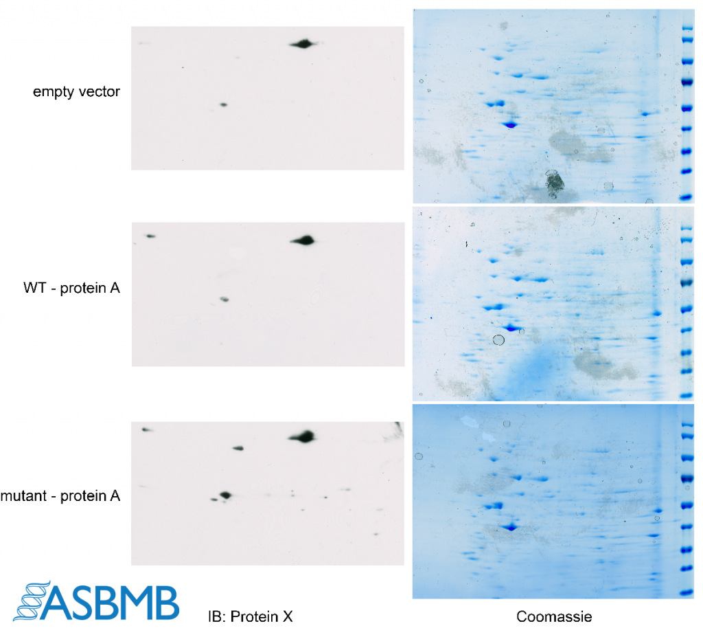
Take a look at the image below, and then take our poll. After that, click on the link below to find out the right answer.

Think you chose the right answer? Click here to find out.
And check out the last Forensics Friday.
Like Retraction Watch? You can make a tax-deductible contribution to support our work, follow us on Twitter, like us on Facebook, add us to your RSS reader, sign up for an email every time there’s a new post (look for the “follow” button at the lower right part of your screen), or subscribe to our daily digest. If you find a retraction that’s not in our database, you can let us know here. For comments or feedback, email us at [email protected].
Perhaps you could alert readers who are using Firefox browser that the multiple choice answer is not visible on this page?
Uuuuumm.. the “duplicated band” does not exist in the original image; only in the solution. Wtf?
The answer key was a little confusing, sorry about that! The circled spots aren’t actually duplicated spots… they were actually erased from the original data. Here’s a link to an image that highlights it a little better: https://twitter.com/KaoruSakabe/status/1135639879675781121. The brush marks are visible when you adjust the levels or brightness/contrast settings.
The gel images also show a suspicious pattern of low resolution bubbles on a high resolution image, and repeating patterns of noise that are duplicated between blots.. could be an innocent explanation I suppose…
Good eye! There are repeating patterns of noise… the Coomassie gels were scanned using a flatbed scanner. They were scanned after being destained in water. The gel was put into a sheet protector before scanning to protect the scanner from water and the same sheet protector was used to scan all the gels. Unfortunately, it looks like the sheet protector was gunky which is why that same background noise is found in all the Coomassie stains.
Yes, but certainly not the identical type stain background on gel 1 and 2 …. that would not happen scanning the gels. No way. Neither if all gels were stained at the same time nor when the gels were scanned after one another.
These types of artifacts are actually common.
This is a good teaching image, with informative comments. (Warning . . . Alert to my anal mindset): Forensically, what is useful about an ‘artifact’ is not that it is – by itself- an ‘unusual’ feature, but that it is one which exists in the same spatial relationship to other features in a separate setting (such as a new scan) where one would not expect that relationship to be quantitatively replicated. In this view, margins of an image can be an ‘artifact.’
Am I seeing things or are the ladders on the Coomassie gels also nearly identical, but with varying degrees of saturation? Ladders are expected to be similar of course, but even down to the individual band angles and tails on each band seems unlikely.