Ever wanted to hone your skills as a scientific sleuth? Now’s your chance.
Thanks to the American Society for Biochemistry and Molecular Biology (ASBMB), which is committed to educating authors on best practices in publishing, figure preparation, and reproducibility, we’re presenting the first of a new series, Forensics Friday.
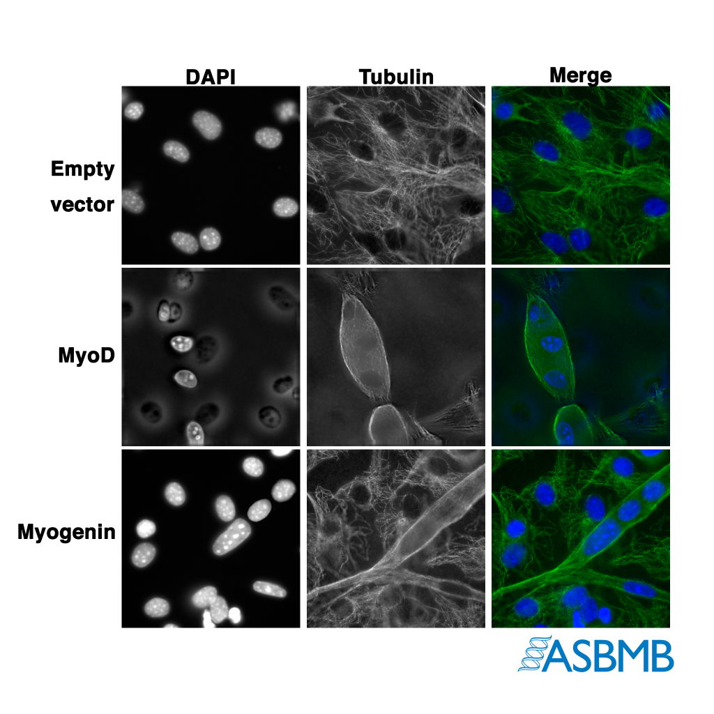
Take a look at the image below, and then take our poll. After that, click on the link below to find out the right answer.

Think you chose the right answer? Click here to find out.
Like Retraction Watch? You can make a tax-deductible contribution to support our growth, follow us on Twitter, like us on Facebook, add us to your RSS reader, sign up for an email every time there’s a new post (look for the “follow” button at the lower right part of your screen), or subscribe to our daily digest. If you find a retraction that’s not in our database, you can let us know here. For comments or feedback, email us at [email protected].
Where is the poll?
Great sports, I love this!
But it’s not only the twin nuclei – the focal depth seems to differ a lot between different parts the myogenin/tubulin image (you can look through a cell and see what’s behind it at 2 o’clock, for example), impossible in a confocal micrograph. This image consists of parts of at least two different ones. I found this easier to notice than the duplications.